
TEGUMENT V10
Evidence spineLinks source imagery, specimen context, calibrated derivatives, review state, and provenance across the workflow.
SEPS uses computation to organize, localize, measure, compare, and audit biological evidence while keeping specimen context and expert authority attached to every claim.
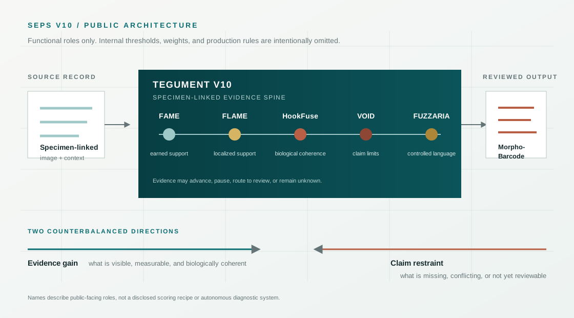
TEGUMENT V10 is the public name for a specimen-linked, fuzzy-first evidence architecture that separates what an image supports, where that support is located, whether the biology is coherent, what limits the claim, and how the result may be described.
This is a public architecture view. Internal thresholds, weights, production settings, transformation details, and unpublished decision logic are intentionally not disclosed.
Preserves raw files, links biological and technical metadata, supports candidate localization, masks, calibration, landmarks, outlines, texture, and versioned derivative outputs.
Asks whether visible structures, life stage, host, infection site, scale, specimen condition, and comparison material make biological sense together.
Determines what the record is allowed to say: archival only, broad-group support, review required, confirmation ready, or unknown.
Acquired images, expert annotations, and derived geometry are displayed as different objects and linked back to the same specimen record.
Each name identifies a function in the evidence chain rather than a claim of autonomous diagnosis. Their separation is intentional: support can rise while the allowable conclusion remains limited.
Links source imagery, specimen context, calibrated derivatives, review state, and provenance across the workflow.
Summarizes how much interpretable support the acquired image contributes to the selected morphology lane.
Tracks whether the supporting cues are attached to a defensible region, structure, or review target.
Reconciles regional image evidence with morphology, life stage, host context, and specimen condition.
Preserves blur, obstruction, missing scale, uncertain orientation, source ambiguity, and other reasons to abstain.
Maps the reviewed evidence state to controlled scientific language and appropriate escalation.
Carries the specimen-linked images, derivatives, measurements, limitations, decisions, and provenance forward.
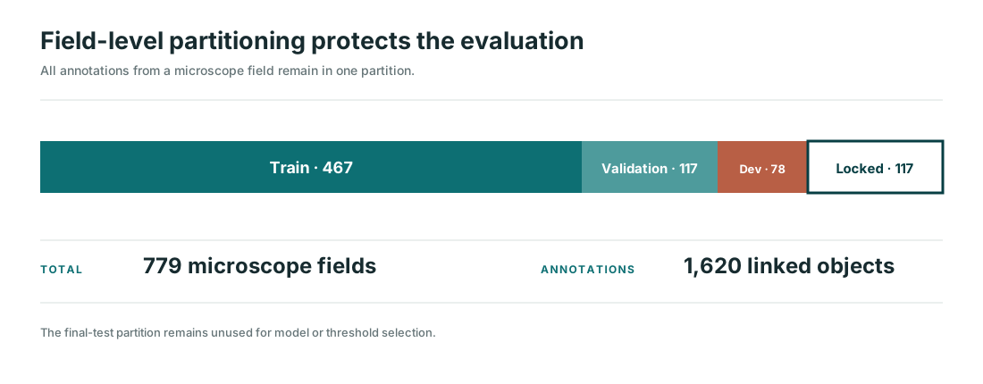
Records are organized as collection event → host → parasite specimen → imaging session → raw image → versioned derivatives. That structure supports traceability, limits pseudoreplication, and makes specimen-level partitioning possible.
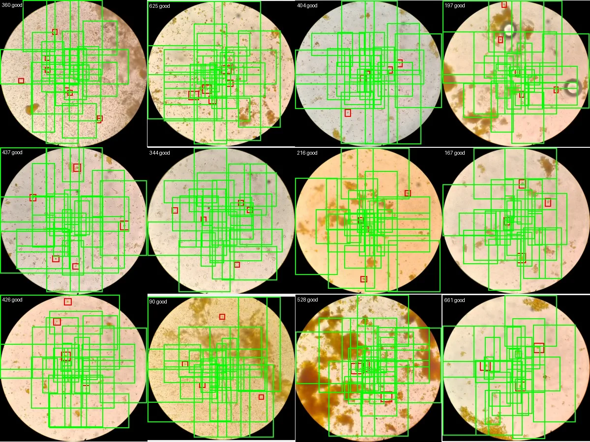
The current detector is a proposal engine. It scans whole microscope fields, ranks candidate regions, and gives later modules a manageable set of places to review. False positives are acceptable at this stage when they reduce the risk of missing a true object.
Centroid capture is therefore the primary developmental target. Boundary overlap, taxonomy, diagnostic interpretation, and reporting language belong to later, separately validated stages.
Exact production settings, unpublished training details, sensitive specimen information, and unreviewed taxonomic conclusions are intentionally omitted from the public site.
Localization recall, mask quality, scale validation, measurement repeatability, shape residuals, calibration, and failure maps.
Specimen-level splits, expert masks, inter-reviewer agreement, difficult negatives, and audit of final reporting language.
New specimens, microscopes, operators, preservation states, laboratories, and out-of-distribution material.