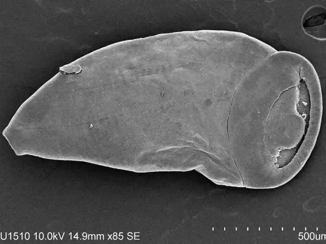
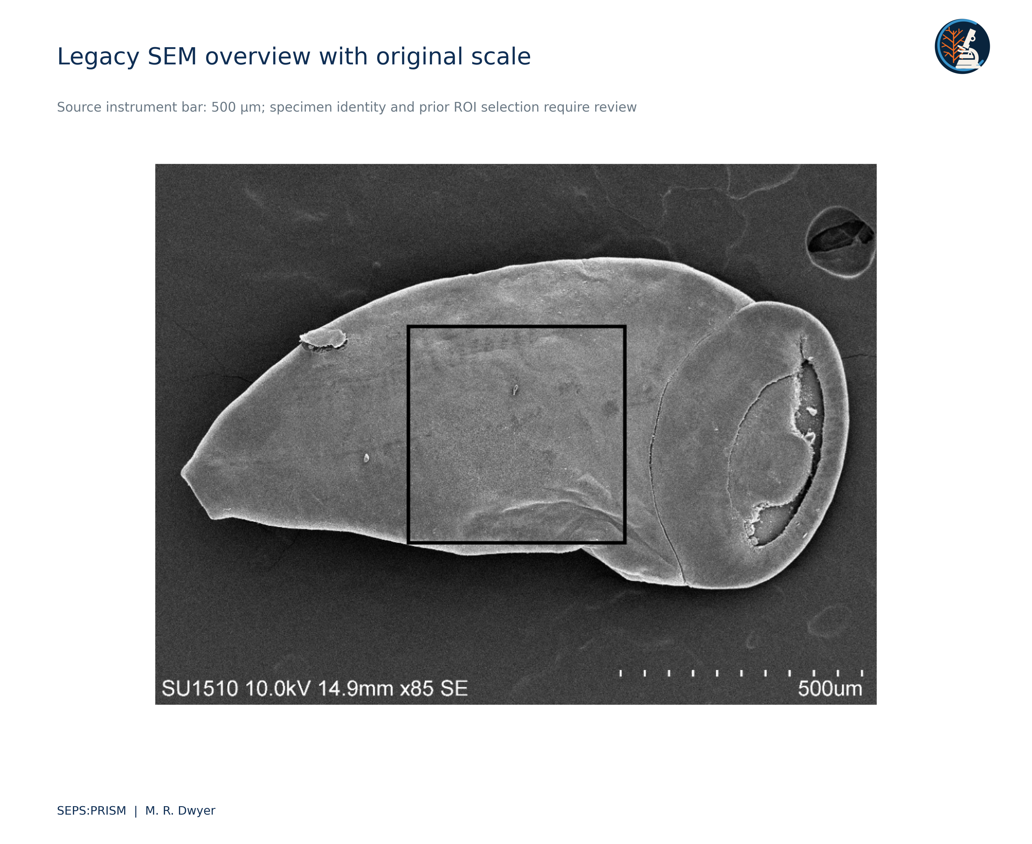
Figure 1 · Trematode under SEMSupplied anatomical annotations shown for illustration; the original instrument labels are retained.SEM imaging: M. en C. María Berenit Mendoza Garfias · Instituto de Biología, Universidad Nacional Autónoma de México (UNAM).
To understand parasite diversity and its place in changing host communities.
SEPS is a nonprofit research organization focused on parasite systematics and disease ecology. Our work connects field observations with comparative morphology, taxonomy, and preserved specimens to investigate neglected and understudied parasites. Computer vision supports this research by organizing images and measurements for expert review.
Building on this foundation, we aim to advance parasite systematics and translate that knowledge into affordable, accessible imaging tools that strengthen diagnostic capacity. Our long-term goal is to develop and validate approaches suited to settings with limited laboratory infrastructure, including those equipped with a smartphone and a microscope, to support reliable parasite identification and efforts to combat parasitic neglected tropical diseases worldwide.
Florida nonprofit organization · U.S. 501(c)(3)
OUR RESEARCH
Parasites, hosts, and the evidence between them.
01
Document diversity
Which parasites occur in sampled hosts, and which anatomical characters support their identification?
02
Resolve host associations
How do parasite records vary among hosts and collection sites? Observed associations provide a starting point for testing transmission and host-switching hypotheses.
03
Test identification methods
Do conventional measurements, geometric morphometrics, and computer vision add reliable identification value when compared with expert morphological review?
OPEN RESEARCH COLLECTION
Explore the evidence behind the work.
Technical reports and proposed laboratory pilots connect parasite systematics with inspectable evidence. Explore documented analyses alongside new research concepts for freshwater snail trematodes and mealworm gregarines.
Field validation, voucher generation, reproducible data products and agency-ready outputs.
THE SEPS METHODOLOGY
SEPS:PRISM
Parasite Recognition through Integrated Specimen Morphology
THE SCIENTIFIC FOUNDATION
A specimen anchors every analysis.
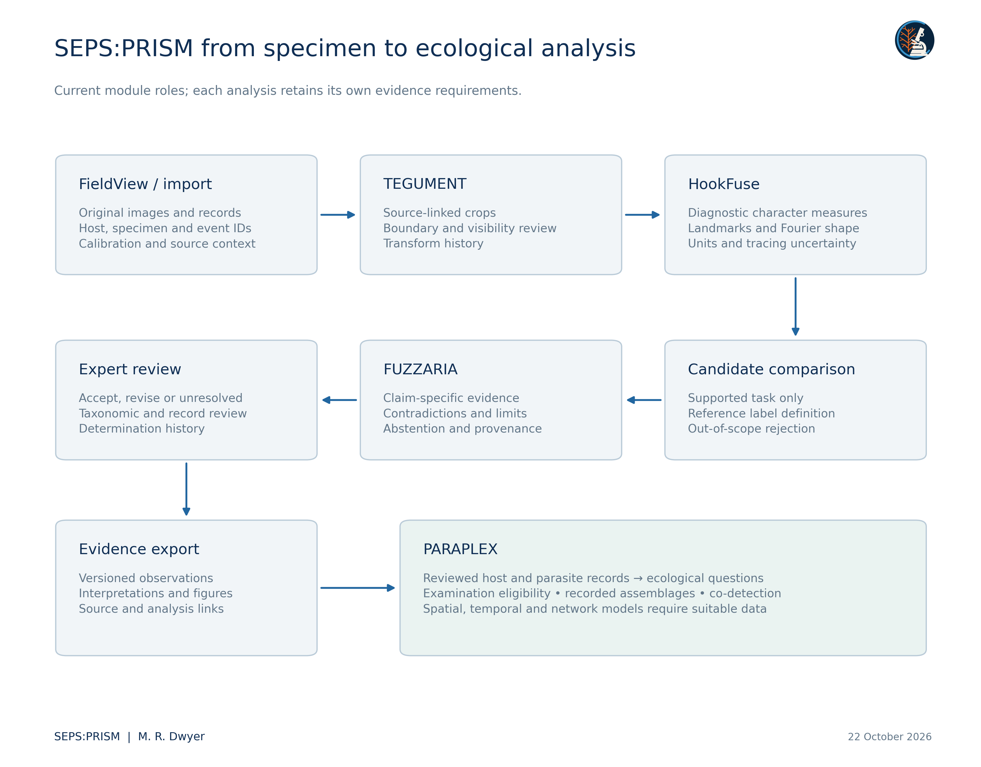
SEPS:PRISM is our integrated methodology for specimen imaging, morphological measurement, and evidence assessment. FieldView supports acquisition, TEGUMENT prepares image evidence, HookFuse examines diagnostic morphology, and FUZZARIA assesses uncertainty and reporting limits. Taxonomic interpretation remains grounded in diagnostic anatomy, reference literature, and expert review.
Each record connects the available images to the host, collection event, preservation history, and voucher. Missing information remains visible. Molecular evidence can be linked when it is available and appropriate to the question.
DEVELOPMENT STATUS
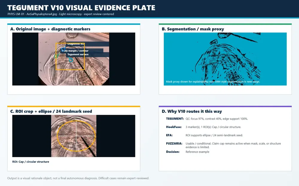
This page describes the SEPS:PRISM methodology and its development requirements. Prototype image-processing changes, proposed robustness pilots, and biological validation are distinct. The workflow diagram does not establish validated automated identification or measured end-to-end performance.
SEPS:PRISM IN PRACTICE
From image to outline.
Explore one specimen photograph, its provisional mask, and a mathematical description of the extracted boundary.
Original imageA trematode SEM photograph from the SEPS archive, retaining its original instrument annotation.SEM imaging: M. en C. María Berenit Mendoza Garfias · Instituto de Biología, Universidad Nacional Autónoma de México (UNAM).
Provisional maskThe apparent outer silhouette is isolated from the background. Boundary placement still needs expert anatomical review.
Fourier approximation20 harmonics approximate the extracted boundary. More harmonics retain more detail of this trace.
FieldView and laboratory uploads enter the same specimen-linked workflow. TEGUMENT is the first processing phase within SEPS:PRISM.
FieldView
Original field photographs, host context, specimen identity, and capture quality.
Laboratory upload
Existing microscopy images, specimen metadata, and calibration where available.
Link the record and select the task
Egg microscopy and adult-worm or hook analysis use separate profiles. Unsupported inputs may remain unresolved.
PHASE 1
TEGUMENT
Segment the specimen and prepare linked image evidence. Review boundaries and retain the original image.
PHASE 2 · WHEN REQUIRED BY THE TASK
HookFuse
Review diagnostic structures, outlines, landmarks, and measurements. Track uncertainty in the contour and its measurements.
PHASE 3
Candidate comparison
Use a supported task model or reference comparison when available. The egg profile bypasses adult-worm and hook extraction.
PHASE 4 · QUALITY CHECKS ALSO APPLY UPSTREAM
FUZZARIA
Assess evidence quality, diagnostic coverage, calibration, provenance, and disagreement. A high model score cannot override missing diagnostic evidence.
PHASE 5
Expert review
Inspect the evidence and record a supported conclusion, a correction request, or an unresolved result.
PHASE 6
Evidence export
Keep the source image, derived views, measurements, quality flags, and review history connected in the specimen record.
Correction path: a reviewer can request a new capture, a corrected mask, or a revised outline. Automated feedback and reprocessing remain proposed work.
Parasite Assemblage and Relationship Analysis across Populations, Landscapes, and Ecological eXchange.
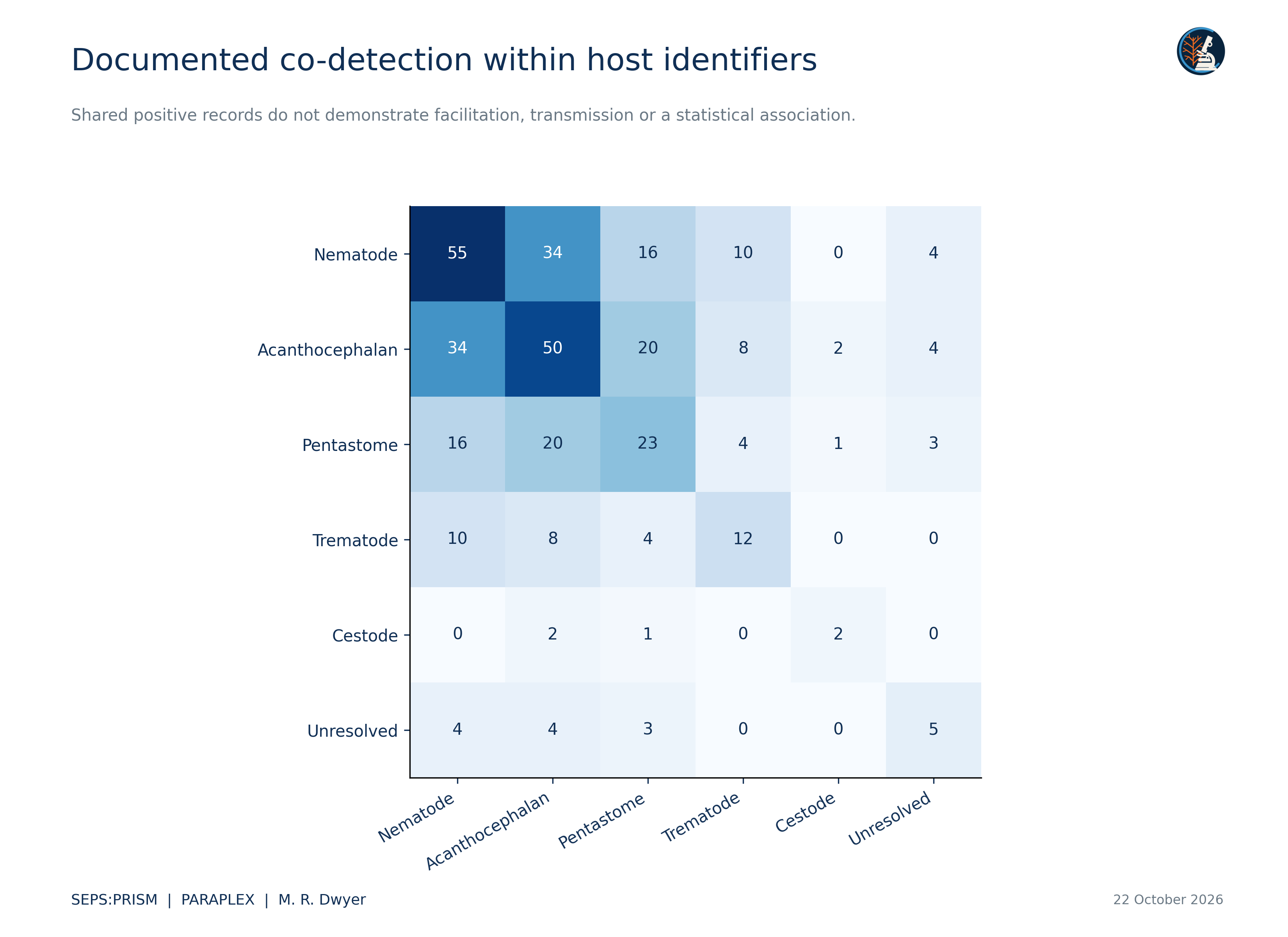
PARAPLEX connects reviewed specimen evidence to host and community questions. The current descriptive analysis preserves the original observation rows, summarizes recorded groups and reconciles candidate links. Population and transmission models require suitable examination records and sampling designs.
These requirements guide development and validation. They describe what must be demonstrated before a feature or identification claim is released.
ACQUISITION REQUIREMENTS
FieldView
Retain originals and failed acquisitions, separate capture failure from no detection, and keep outputs linked to the current image. Offline recovery and delayed or stale outputs require device testing.
PROTOTYPE CHANGES AND PROPOSED PILOTS
TEGUMENT
Reported whole-worm prototype changes separate specimen outlines and confine internal contrast patches to their selected outlines. Boundary preservation and thin-structure continuity need anatomical review; contrast patches are not confirmed anatomy.
MORPHOLOGY REQUIREMENTS
HookFuse
Preserve fine attachment structures, review contour uncertainty, and carry that uncertainty into measurements. Shape comparison requires comparable views and reviewed traces. Retrieved look-alike images support expert inspection rather than establish identity.
EVIDENCE AND RELEASE REQUIREMENTS
FUZZARIA
Keep quality dimensions and contradictory evidence visible. Assess boundary and anatomical errors separately from mask overlap. Unsupported inputs can be rejected; an unresolved outcome is acceptable. Restored or generated anatomical detail is inadmissible identification evidence.
Spatial coverage for selecting review examples, constrained thin-structure tracing, measurement-uncertainty propagation, and FieldView response-time checks remain proposed pilots until tested on relevant SEPS specimens or devices. The methodology does not treat those pilots as completed validation.
WHAT EACH ANALYSIS CONTRIBUTES
Methods with an explicit purpose.
Comparative morphology and conventional morphometrics
Diagnostic anatomy is compared with taxonomic descriptions, keys, and appropriate reference specimens. Calibrated lengths, widths, angles, and ratios are recorded where the structures and views support them. Hook geometry and other attachment structures are a focused application within HookFuse.
Measurements retain their units, calibration source, view, and reviewer. Missing calibration prevents claims about absolute physical size. Incomplete anatomy or conflicting metadata limits the taxonomic conclusion.
Geometric morphometrics and outline analysis
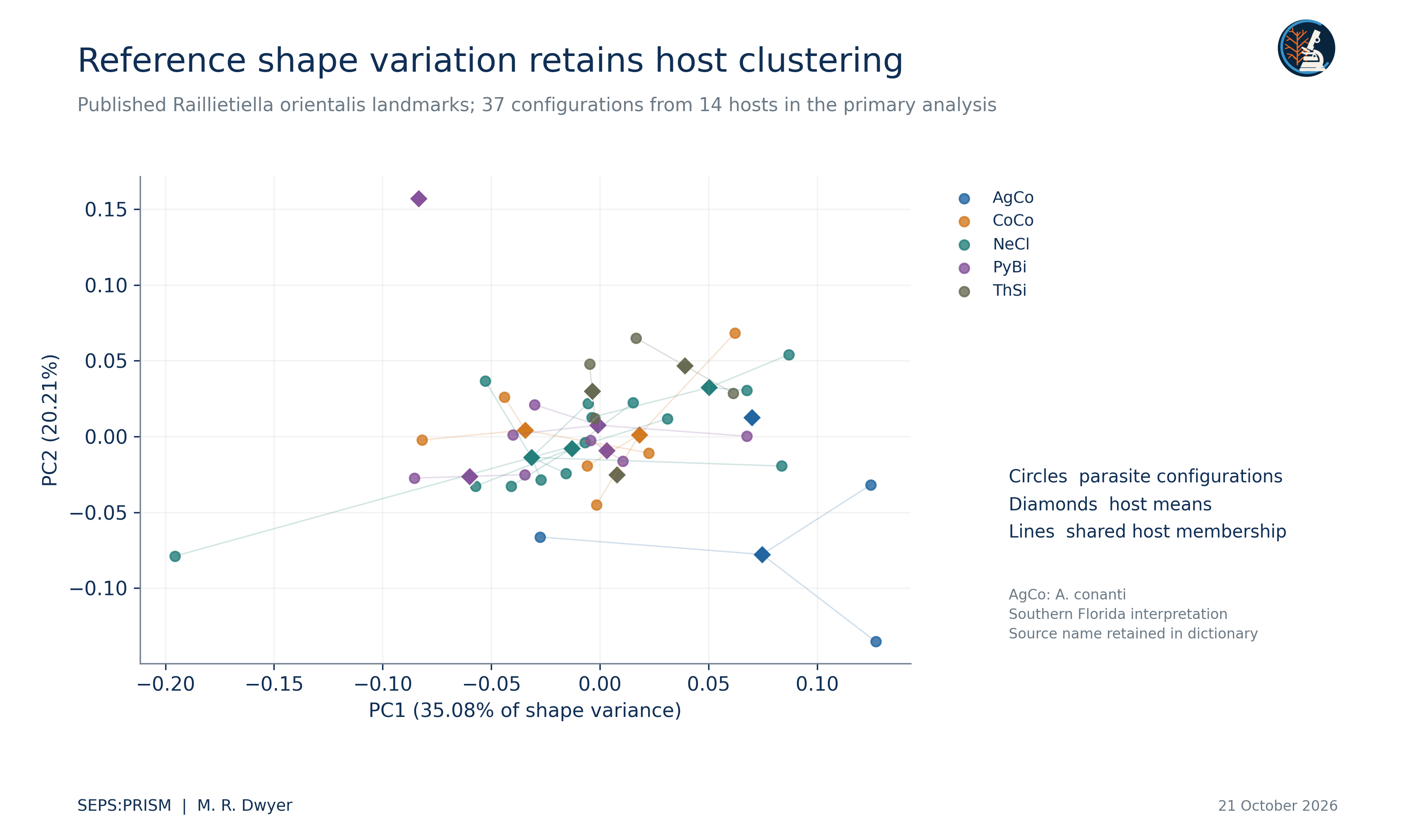
The proposed extension uses anatomically corresponding landmarks and reviewed semilandmarks to compare shape. Generalized Procrustes alignment removes differences in position, rotation, and overall scale; size is retained separately so allometry can be investigated.
Principal component analysis can describe shape variation without establishing species identity. Elliptic Fourier analysis is an optional branch for suitable closed outlines. View, orientation, preservation, and tracing error must be controlled before comparisons are interpreted.
Computer vision and Vision Transformers
TEGUMENT organizes conservative image preparation, segmentation, and diagnostic-region review. Masks must preserve the anatomy needed for identification and measurement. Original photographs remain available alongside every processed derivative.
Vision Transformers are a proposed computer-vision extension for learning image features and testing candidate classification or reference-image comparison. Attention maps alone do not establish an anatomical explanation. Model version, reference data, preprocessing, and review status accompany any experimental output.
FUZZARIA and expert review
FUZZARIA organizes evidence adequacy and uncertainty to prioritize review. The proposed integration retains separate assessments for image quality, diagnostic coverage, calibration, and provenance, with explicit notes when analyses disagree.
Insufficient evidence can lead to recapture, further examination, or an unresolved record. No-detection images and inadequate acquisitions are retained with distinct outcomes. A model detecting no target is not, by itself, evidence that a host is uninfected.
PROPOSED VALIDATION
Does each added method improve identification?
The pilot compares conventional morphology and measurements with the added value of geometric morphometrics, Vision Transformers, and their combination. Expert-reviewed references are required before performance is reported.
Independent evaluation
Repeated images of a specimen remain in one data partition. Host and collection grouping are respected where required by the study design.
Separate endpoints
Detection, measurement error, shape repeatability, and taxonomic classification are evaluated separately. Unresolved cases remain visible.
COMPONENTS WITHIN SEPS:PRISM
Tools that serve the specimen.
SEPS:PRISM names the complete methodology. FieldView documents acquisition, TEGUMENT prepares visual evidence, HookFuse organizes diagnostic characters, and FUZZARIA supports review. Lens is the separate mobile image-readiness interface, not an extra processing phase.
Image preview only. No record is saved and no file is uploaded. Use the documentation template to prepare a linked record.
VISUAL RESEARCH ARCHIVE
Archive
Original views, diagnostic details, and linked documentation.
These are image entries. Physical specimen identity, host association, and voucher linkage require verification. R-001, R-006, and R-017 now open structured draft records; their missing fields are shown explicitly.
This collection documents completed calculations, comparative microscopy and specimen-linked methods. These author-reviewed technical and educational reports are not peer-reviewed journal publications or a validated clinical diagnostic system.
M. R. Dwyer · Updated 23 October 2026 · 6 technical reports · 2 proposed pilots · 28 figure sets
This proposed pilot would build a small reference collection of larval trematodes from freshwater snails using suitable preserved material. Microscopy and specimen records would document recognizable forms. Selected DNA markers would then test identification where preservation permits. The intended result is an illustrated reference that connects each identification to its specimen, images and supporting evidence.
This proposed pilot would examine naturally occurring gregarines in commercially reared mealworms. It would ask how parasite shape and developmental stage relate to molecular identity. Photographs and measurements would be linked to selected DNA sequences where feasible. The intended outcome is a reference atlas that documents variation and supports careful identification of these overlooked invertebrate parasites.
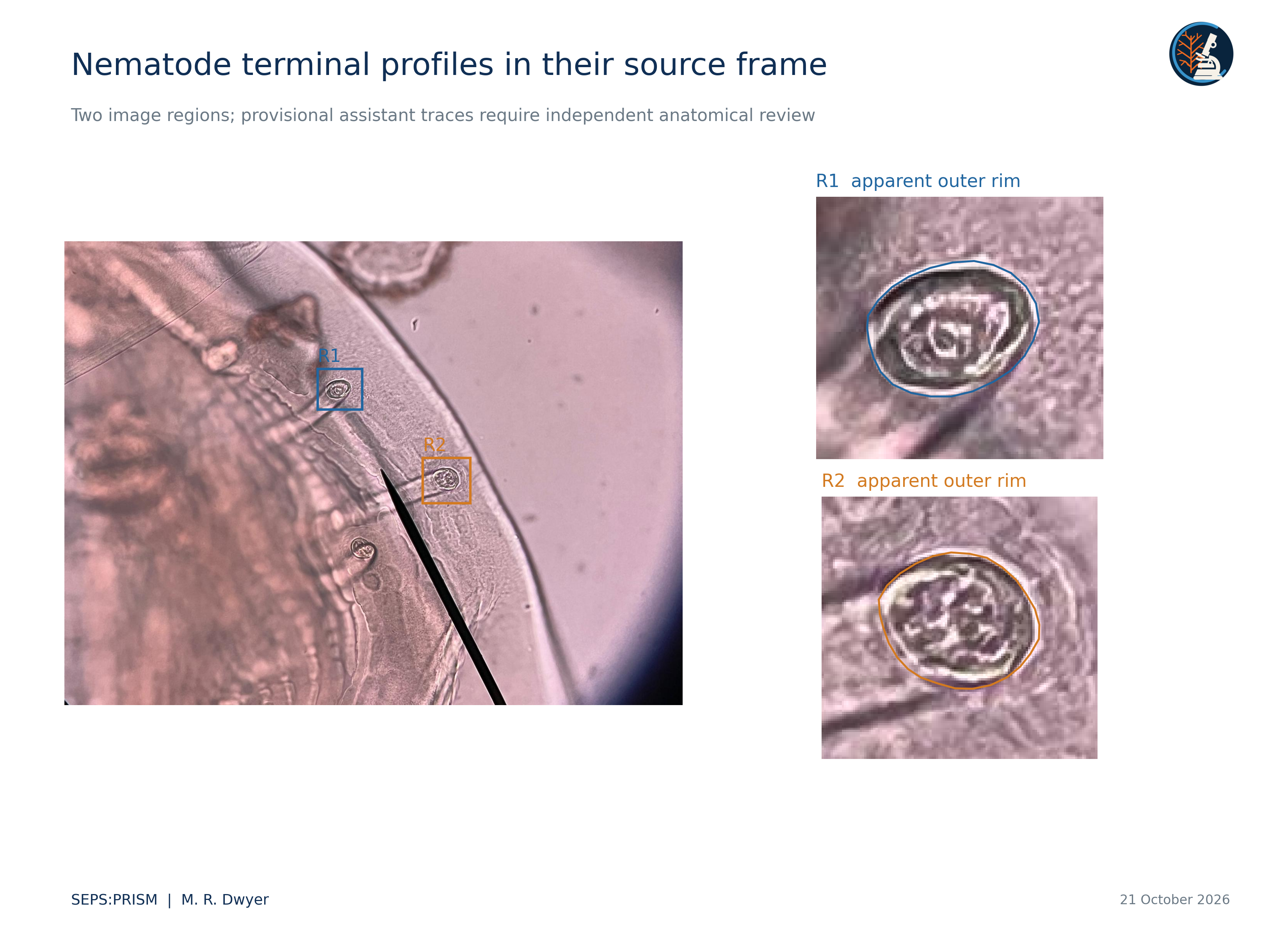
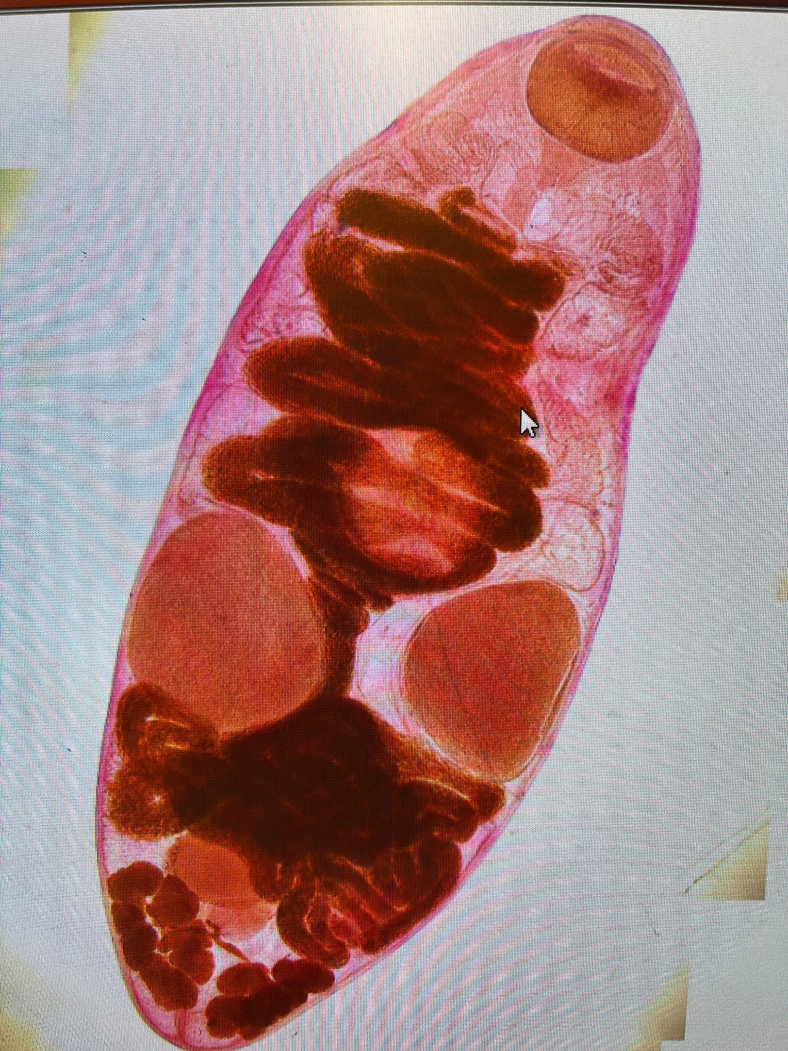
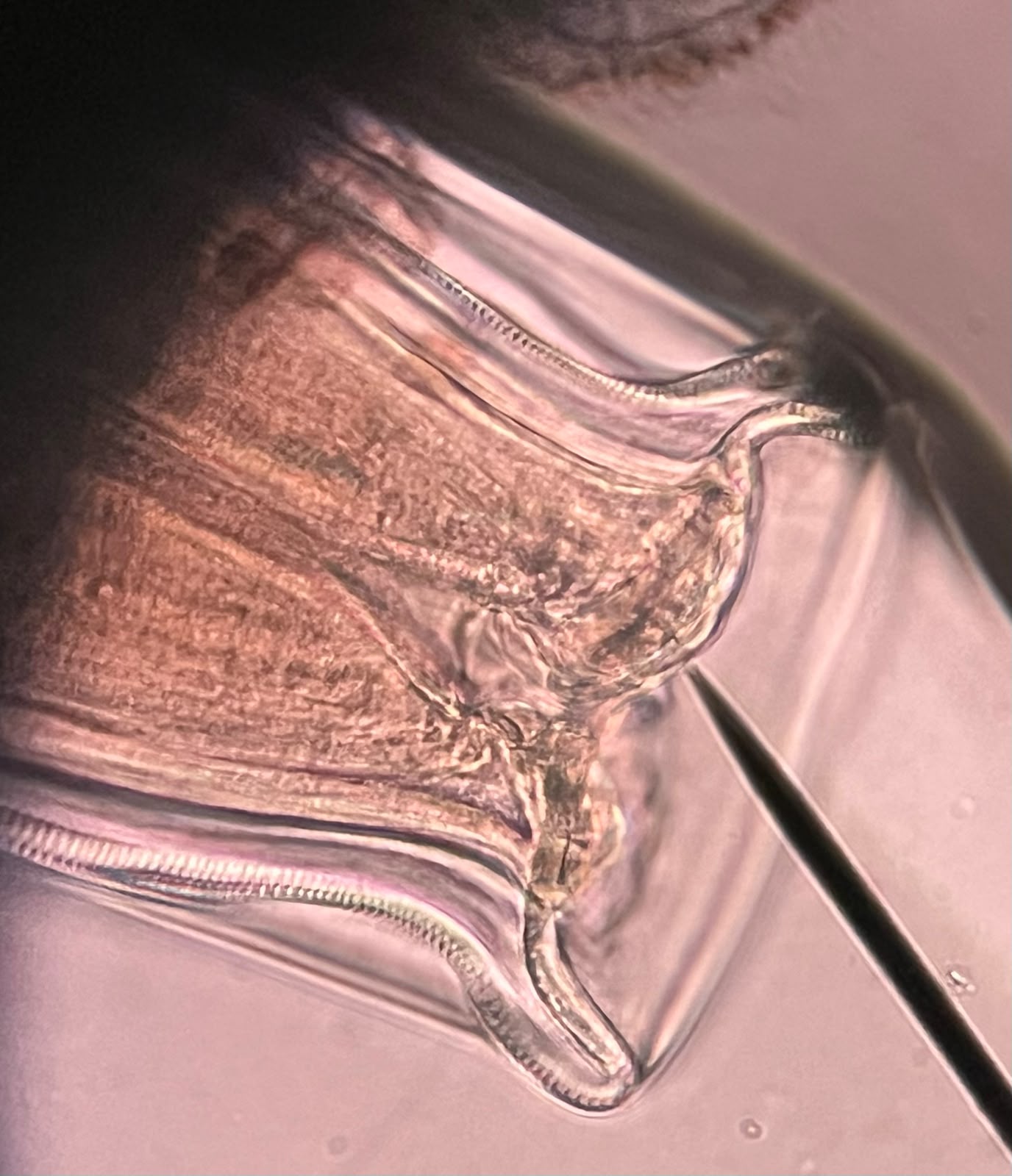
Whole trematode specimen, cleared with ethanol and stained with paracarmine. Brightfield microscopy, 10× magnification.Nematode buccal capsule, cleared in glycerin, with the cuticle and oesophagus visible in detail. Brightfield microscopy, 40× magnification.
SEPS
Society for Evolutionary Parasite Systematics
U.S. 501(c)(3) nonprofit
Parasite diversity. Documented carefully.
SEPS is a U.S. 501(c)(3) nonprofit research organization focused on parasite systematics and disease ecology. Comparative morphology, taxonomy, ecology, and preserved specimens guide our work on neglected and understudied parasites. Computer vision and quantitative shape analysis support these scientific questions.
Building on this foundation, we aim to advance parasite systematics and translate that knowledge into affordable, accessible imaging tools that strengthen diagnostic capacity. Our long-term goal is to develop and validate approaches suited to settings with limited laboratory infrastructure, including those equipped with a smartphone and a microscope, to support reliable parasite identification and efforts to combat parasitic neglected tropical diseases worldwide.